lv apical aneurysm | american heart association guidelines 2024 lv apical aneurysm A ventricular aneurysm is a bulge or weakened area in the wall of your heart’s ventricles. Learn about the types, causes, complications and treatments of ventri. $14.99
0 · left ventricular aneurysm vs pseudoaneurysm
1 · left ventricular aneurysm repair surgery
2 · american heart association guidelines 2024
3 · aha heart failure guidelines 2024
4 · Lv pseudoaneurysm vs true aneurysm
5 · Lv apical aneurysm management
6 · Lv aneurysm vs pseudoaneurysm echo
7 · Lv aneurysm on echo
$259.99
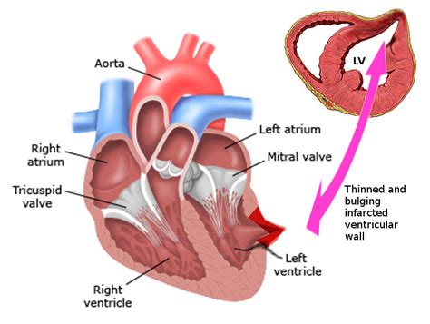
A ventricular aneurysm is a bulge or weakened area in the wall of your heart’s ventricles. Learn about the types, causes, complications and treatments of ventri. A significant left ventricular (LV) aneurysm is present in 30% to 35% of acute transmural myocardial infarction. The two major risk factors for developing LV aneurysm include total occlusion of the left anterior descending artery .A ventricular aneurysm is a bulge or weakened area in the wall of your heart’s ventricles (lower pumping chambers). It most commonly occurs in the left ventricle after a heart attack causes heart muscle to die or weaken. Left ventricular aneurysm formation following acute STEMI causes persistent ST elevation on the ECG. ECG Features of Left Ventricular Aneurysm. ST elevation seen > 2 weeks following an acute myocardial infarction. Most commonly seen in the precordial leads. May exhibit concave or convex morphology. Usually associated with well-formed Q- or QS waves
Left ventricular (LV) aneurysms and pseudoaneurysms are two complications of myocardial infarction (MI) that can lead to death or significant morbidity. This topic reviews the diagnosis and management of patients with aneurysms or pseudoaneurysms caused by MI. Apical aneurysms were diagnosed on preoperative transthoracic echocardiography, on cardiac magnetic resonance imaging, or by visual inspection of the left ventricular apex at operation. The mean age of patients undergoing operation for apical aneurysms was 56±14 years, and 52% were men.
The left ventricular (LV) ejection fraction was 30% and an apical aneurysm could be clearly identified. The preoperative transthoracic echocardiogram without and with intravenous contrast confirmed a 3.5x 1.5 cm left ventricular aneurysm.Left ventricular (LV) apical aneurysms in hypertrophic cardiomyopathy (HCM) are associated with sudden cardiac death (SCD), formation of apical thrombus and thromboembolic events, and progression to end-stage disease.
left ventricular aneurysm vs pseudoaneurysm

Is anticoagulation really indicated for laminated thrombus (not a more mobile, round, mural thrombus)? 7. Is DOAC a reasonable alternative to warfarin for the prevention and treatment of LV thrombus? 8. What management options are there in patients with persistent LV thrombus despite therapy?A previously under-recognized subset of hypertrophic cardiomyopathy (HCM) patients with left ventricular (LV) apical aneurysms is being identified with increasing frequency. However, risks associated with this subgroup are unknown.Background: Left ventricular (LV) apical aneurysms in hypertrophic cardiomyopathy (HCM) are a recognized risk marker for adverse cardiovascular events. There is variable practice among clinicians and discordance between international guidelines regarding treatment recommendations and prognostication for this important phenotype.
A significant left ventricular (LV) aneurysm is present in 30% to 35% of acute transmural myocardial infarction. The two major risk factors for developing LV aneurysm include total occlusion of the left anterior descending artery .
A ventricular aneurysm is a bulge or weakened area in the wall of your heart’s ventricles (lower pumping chambers). It most commonly occurs in the left ventricle after a heart attack causes heart muscle to die or weaken. Left ventricular aneurysm formation following acute STEMI causes persistent ST elevation on the ECG. ECG Features of Left Ventricular Aneurysm. ST elevation seen > 2 weeks following an acute myocardial infarction. Most commonly seen in the precordial leads. May exhibit concave or convex morphology. Usually associated with well-formed Q- or QS waves Left ventricular (LV) aneurysms and pseudoaneurysms are two complications of myocardial infarction (MI) that can lead to death or significant morbidity. This topic reviews the diagnosis and management of patients with aneurysms or pseudoaneurysms caused by MI. Apical aneurysms were diagnosed on preoperative transthoracic echocardiography, on cardiac magnetic resonance imaging, or by visual inspection of the left ventricular apex at operation. The mean age of patients undergoing operation for apical aneurysms was 56±14 years, and 52% were men.
The left ventricular (LV) ejection fraction was 30% and an apical aneurysm could be clearly identified. The preoperative transthoracic echocardiogram without and with intravenous contrast confirmed a 3.5x 1.5 cm left ventricular aneurysm.Left ventricular (LV) apical aneurysms in hypertrophic cardiomyopathy (HCM) are associated with sudden cardiac death (SCD), formation of apical thrombus and thromboembolic events, and progression to end-stage disease. Is anticoagulation really indicated for laminated thrombus (not a more mobile, round, mural thrombus)? 7. Is DOAC a reasonable alternative to warfarin for the prevention and treatment of LV thrombus? 8. What management options are there in patients with persistent LV thrombus despite therapy?
A previously under-recognized subset of hypertrophic cardiomyopathy (HCM) patients with left ventricular (LV) apical aneurysms is being identified with increasing frequency. However, risks associated with this subgroup are unknown.
left ventricular aneurysm repair surgery

dior cherry 015
american heart association guidelines 2024
Agnès Maltais, Québec. Députée de Taschereau Porte-parole de l’opposition officielle: -en matière de laïcité -responsable de la région de la Capitale-Nationale
lv apical aneurysm|american heart association guidelines 2024


